Something Powerful

Tell The Reader More

The headline and subheader tells us what you're offering, and the form header closes the deal. Over here you can explain why your offer is so great it's worth filling out a form for.

Remember:

  • Bullets are great
  • For spelling out benefits and
  • Turning visitors into leads.

DiversityNursing Blog

Breast-friendly, radiation-free alternative to mammograms in the works

Posted by Pat Magrath

Tue, Nov 15, 2016 @ 10:38 AM

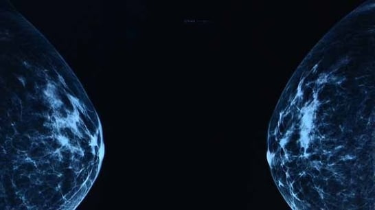
Mammogram-hero.jpgMammograms, we know how barbaric and uncomfortable the procedure is for all of us. I often find myself thinking, there’s got to be a better way. Wouldn’t it be great if we had something that gives a better picture, doesn’t use radiation, and doesn’t flatten our breasts during the process? We’d all like to skip our mammograms entirely, but the prospect of early cancer detection keeps us going back year after year.
f08f51fe01cce526ba77fe35ebcd9ae2.jpg
Well, it looks like some progress is being made in the world using sound waves. Check out this article and let us know your thoughts.

Each year, millions of women undergo mammograms for early detection of possible breast cancer. It's an unpleasant procedure that uses X-rays. Researchers at TU Eindhoven are working on a 'breast-friendly' method, without radiation, that is more accurate and generates 3D rather than 2D images. They published their proof of concept earlier this month in the online journal Scientific Reports.

In the regular screening method the breast is squeezed tight between two plates in order to produce one or more good X-ray photos. Apart from being unpleasant, it is not without risk. The X-rays used can themselves be a contributor to the onset of cancer. Moreover, it is often unclear whether the anomaly found is malignant lesion or not. More than two-thirds of the cases where something worrying can be seen on the X-ray photos is a false-positive: after biopsies, they are not found to be cancer. This is why science is seeking alternatives.

Researchers at TU Eindhoven have now cleared a major scientific hurdle towards a new technology in which the patient lies on a table and the breast hangs freely in a bowl. Using special echography (inaudible sound waves) a 3D image is made of the breast. Any cancer is clearly identifiable on the generated images; the researchers therefore expect there to be many fewer false-positive results.

The new technology builds on the patient-friendly prostate cancer detection method developed at TU/e whereby the doctor injects the patient with harmless microbubbles. An echoscanner allows these bubbles to be precisely monitored as they flow through the blood vessels of the prostate. Since cancer growth is associated with the formation of chaotic microvessels, the presence and location of cancer become visible. This method works well for the prostate and this is now being widely tested in hospitals in the Netherlands, China and, soon, Germany. For breast cancer the method had not yet been suitable because the breast shows excessive movement and size for accurate imaging by standard echography.

Researchers Libertario Demi, Ruud van Sloun and Massimo Mischi have now developed a variant of the echography method that is suitable for breast investigation. The method is known as Dynamic Contrast Specific Ultrasound Tomography. Echography with microbubbles uses the fact that the bubbles will vibrate in the blood at the same frequency as the sound produced by the echoscanner, as well as at twice that frequency; the so-called second harmonic. By capturing the vibration, you know where the bubbles are located. But body tissue also generates harmonics, and that disturbs the observation.

For the new method the researchers are using a phenomenon that Mischi happened upon by chance and later investigated its properties together with Demi. They saw that the second harmonic was a little delayed by the gas bubbles. The researchers have now developed a new visualization method. The more bubbles are encountered by the sound on its route, the bigger the delay compared to the original sound. By measuring this delay, the researchers can thus localize the air bubbles and do so without any disturbance because the harmonic generated by the body tissue is not delayed, and is therefore discernible. This difference, however, can only be seen if the sound is captured on the other side. So this method is perfectly suited to organs that can be approached from two sides, like the breast.

The researchers are currently putting together an international, strong medical team to start performing preclinical studies. Application in practice is certainly ten or so years away, Mischi expects. Moreover, he forecasts that the technology that has been developed will probably not operate on a standalone basis but in combination with other methods, which will create a better visualization. One of the candidates for this elastography, a variant of echography whereby the difference in the rigidity of the tumor and healthy tissue can be used to detect cancer.

New Call-to-action

Topics: mammography, breast screening, mammograms

New Device May Ease Mammography Discomfort

Posted by Erica Bettencourt

Wed, Dec 03, 2014 @ 12:17 PM

141125074824 large resized 600

Researchers have developed a new device that may result in more comfortable mammography for women. According to a study being presented next week at the annual meeting of the Radiological Society of North America (RSNA), standardizing the pressure applied in mammography would reduce pain associated with breast compression without sacrificing image quality.

Compression of the breast is necessary in mammography to optimize image quality and minimize absorbed radiation dose. However, mechanical compression of the breast in mammography often causes discomfort and pain and deters some women from mammography screening.

An additional problem associated with compression is the variation that occurs when the technologist adjusts compression force to breast size, composition, skin tautness and pain tolerance. Over-compression, or unnecessarily high pressures during compression, is common in certain European countries, especially for women with small breasts. Over-compression occurs less frequently in the United States, where under-compression, or extremely low applied pressure, is more common.

"This means that the breast may be almost not compressed at all, which increases the risks of image quality degradation and extra radiation dose," said Woutjan Branderhorst, Ph.D., researcher in the Department of Biomedical Engineering and Physics at the Academic Medical Center in Amsterdam.

Overall, adjustments in force can lead to substantial variation in the amount of pressure applied to the breast, ranging from less than 3 kilopascals (kPa) to greater than 30 kPa.

Dr. Branderhorst and colleagues theorized that a compression protocol based on pressure rather than force would reduce the pain and variability associated with the current force-based compression protocol. Force is the total impact of one object on another, whereas pressure is the ratio of force to the area over which it is applied.

The researchers developed a device that displays the average pressure during compression and studied its effects in a double-blinded, randomized control trial on 433 asymptomatic women scheduled for screening mammography.

Three of the four compressions for each participant were standardized to a target force of 14 dekanewtons (daN). One randomly assigned compression was standardized to a target pressure of 10 kPa.

Participants scored pain on a numerical rating scale, and three experienced breast screening radiologists indicated which images required a retake. The 10 kPa pressure did not compromise radiation dose or image quality, and, on average, the women reported it to be less painful than the 14 daN force.

The study's implications are potentially significant, Dr. Branderhorst said. There are an estimated 39 million mammography exams performed every year in the U.S. alone, which translates into more than 156 million compressions. Pressure standardization could help avoid a large amount of unnecessary pain and optimize radiation dose without adversely affecting image quality or the proportion of required retakes.

"Standardizing the applied pressure would reduce both over- and under-compression and lead to a more reproducible imaging procedure with less pain," Dr. Branderhorst said.

The device that displays average pressure is easily added to existing mammography systems, according to Dr. Branderhorst.

"Essentially, what is needed is the measurement of the contact area with the breast, which then is combined with the measured applied force to determine the average pressure in the breast," he said. "A relatively small upgrade of the compression paddle is sufficient."

Further research will be needed to determine if the 10 kPa pressure is the optimal target.

The researchers are also working on new methods to help mammography technologists improve compression through better positioning of the breast.

Source: www.sciencedaily.com

Topics: mammography, tests, screenings, technology, health, healthcare, nurses, doctors, medical, breast cancer

Recent Jobs

Article or Blog Submissions

If you are interested in submitting content for our Blog, please ensure it fits the criteria below:
  • Relevant information for Nurses
  • Does NOT promote a product
  • Informative about Diversity, Inclusion & Cultural Competence

Agreement to publish on our DiversityNursing.com Blog is at our sole discretion.

Thank you

Subscribe to Email our eNewsletter

Recent Posts

Posts by Topic

see all